Sketch And Label Of A Cross Section Of A Long Bone / The outside of a bone is covered in a thin layer of dense irregular connective tissue called the periosteum.
Sketch And Label Of A Cross Section Of A Long Bone / The outside of a bone is covered in a thin layer of dense irregular connective tissue called the periosteum.. Cartilaginous area at the ends of long bones where lengthwise growth takes place in the immature skeleton. Cross section of a long bone. While it is not as hard as compact bone, spongy bone plays an important role of protecting the marrow where blood cells are produced. Click on the tags below to find other quizzes on the same subject. Shannan muskopf october 16, 2020.
Plates of cartilage, also known as growth plates which allow the long bones to grow during childhood. Forms the larger rounded ends of long bones. Continue to label this drawing as you explore the inside of the bone. Smartdraw includes 1000s of professional healthcare and anatomy chart templates that you can modify and make your own. Cow and human long bones have a similar general structure.
Anatomy And Physiology Of Animals The Skeleton Wikibooks Open Books For An Open World from upload.wikimedia.org The end of a growing tibia, cut lengthwise*. Bone matrix and cells bone matrix osseous tissue is a connective tissue and like all connective tissues contains relatively few cells and large amounts of extracellular matrix. The diaphysis is the tubular shaft that runs between the proximal and distal ends of the bone. Once we stop growing (between 18. Learners should accurately draw a long bone, resembling that in figure 6.24. The diaphysis of a long bone is composed of bone tissue while the epiphysis is made of bone tissue. There is a printable worksheet available for download here so you can take the quiz with pen and paper. Draw and label the following structures as they appear using the 10x objective o bone marrow o bony trabeculae activity 5.2.3:
Bone matrix and cells bone matrix osseous tissue is a connective tissue and like all connective tissues contains relatively few cells and large amounts of extracellular matrix.
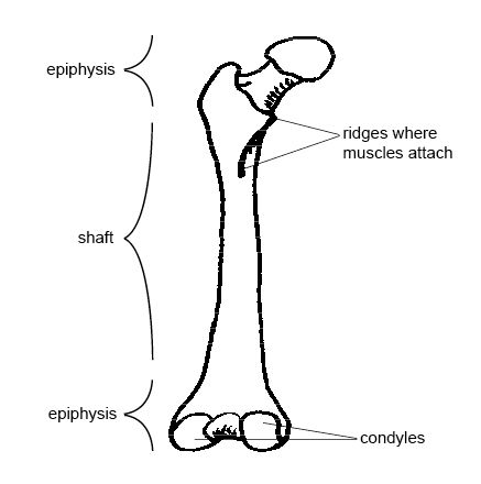
The diaphysis of a long bone is composed of bone tissue while the epiphysis is made of bone tissue. In these labeled examples, a human femur is represented without identifying many of the unique characteristics that help differentiate the femur bone from other bones in the human body. This is an online quiz called label the long bone. Make sure learners follow all the criteria for a biological drawing. The head of each end of a long bone consists largely of spongy bone and is covered with hyaline cartilage. Marks should be deducted for shading or colouring. It suggests that the bone will have equal strength in all directions. Once we stop growing (between 18. Area between the diaphysis and epiphysis at both ends of the bone. On this page, you will find two images i created that illustrate the parts of a long bone and long bone structure. Cartilaginous area at the ends of long bones where lengthwise growth takes place in the immature skeleton. Use colored pencils to draw and label the following structures as they appear using the 40x objective, or by looking at an image from the internet. A section view is a view used on a drawing to show an area or hidden part of an object by cutting away or removing some of that object.
Cartilaginous area at the ends of long bones where lengthwise growth takes place in the immature skeleton. Anatomy of a long bone 1. Marks should be deducted for shading or colouring. Diaphysis • shaft of the long bone. Cross section of a long bone.
Sketch And Label Of A Cross Section Of A Long Bone Cross Section Of Right Kidney Photograph By Science Source from i1.wp.com Foot bone anatomy x ray 12 photos of the foot bone anatomy x ray foot bone anatomy x ray, bone, foot bone anatomy x ray. End of a long bone. Draw labelled diagram showing relations of label the parts of a long bone. Cow and human long bones have a similar general structure. Continue to label this drawing as you explore the inside of the bone. The structure of a long bone consists of several sections:. Draw and label the following structures as they appear using the 10x objective o bone marrow o bony trabeculae activity 5.2.3: There is a printable worksheet available for download here so you can take the quiz with pen and paper.
(do not copy and paste a picture from the text or internet.)
Foot bone anatomy x ray 12 photos of the foot bone anatomy x ray foot bone anatomy x ray, bone, foot bone anatomy x ray. Make sure learners follow all the criteria for a biological drawing. Label the haversian canal, osteocyte (mature bone cell) in lacuna, and canaliculi. Make a pencil sketch and use markers or colored pencils to add details. An osteon is the basic functional and structural unit of mature compact bone. A long bone has two parts: The diaphysis is the tubular shaft that runs between the proximal and distal ends of the bone. Use colored pencils to draw and label the following structures as they appear using the 40x objective, or by looking at an image from the internet. Only the bottom portion of this bone extends as far as the hoof capsule. The structure of a long bone allows for the best visualization of all of the parts of a bone ( figure 6.7 ). Label lines should not cross ; Smartdraw includes 1000s of professional healthcare and anatomy chart templates that you can modify and make your own. This is the long central shaft.
The digital cushion sits just behind the pedal bone and above the sensitive frog. It is located between the elbow joint and the shoulder. Only the bottom portion of this bone extends as far as the hoof capsule. Shannan muskopf october 16, 2020. The diaphysis is the tubular shaft that runs between the proximal and distal ends of the bone.
Sketch And Label Of A Cross Section Of A Long Bone Cross Section Of Right Kidney Photograph By Science Source from i0.wp.com Anatomy of a long bone 1. Foot bone anatomy x ray 12 photos of the foot bone anatomy x ray foot bone anatomy x ray, bone, foot bone anatomy x ray. A section view is a view used on a drawing to show an area or hidden part of an object by cutting away or removing some of that object. A long bone is a bone that has greater length than width. This is an online quiz called long bone anatomy. Draw labelled diagram showing relations of label the parts of a long bone. In the space provided draw a longitudinal section of a long bone and label the following parte proximal epiphysis, distal epiphysis, diaphysis, metaphysis, medullary cavity, epiphyseal line 2. While it is not as hard as compact bone, spongy bone plays an important role of protecting the marrow where blood cells are produced.
(do not copy and paste a picture from the text or internet.)
The head of each end of a long bone consists largely of spongy bone and is covered with hyaline cartilage. A typical long bone shows the gross anatomical characteristics of bone. Area between the diaphysis and epiphysis at both ends of the bone. Then, fill in the table below to describe each. Make sure learners follow all the criteria for a biological drawing. Cow and human long bones have a similar general structure. The digital cushion sits just behind the pedal bone and above the sensitive frog. The diaphysis and the epiphysis. What is a section view ? Label lines should not cross ; Unit 3 part 1 x section bone. Use colored pencils to draw and label the following structures as they appear using the 40x objective, or by looking at an image from the internet. Also known as the middle phalanx, the short pastern bone sits on top of the articulating joint of the pedal bone and underneath the long pastern bone.


0 Komentar